X-rays & 3D imaging
Digital images with low radiation, including 3D cone beam CT (CBCT).
What is X-rays & 3D imaging?
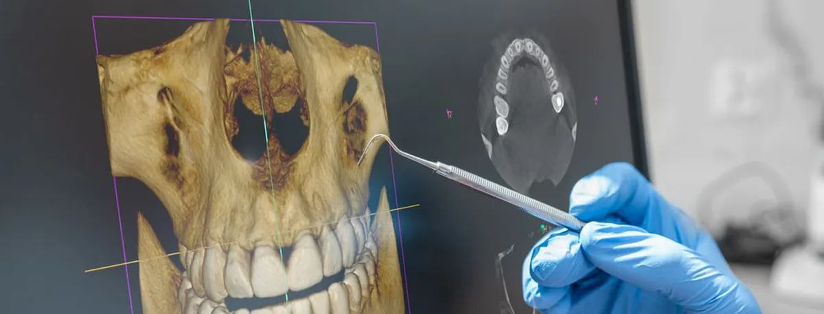
Digital X-rays show what cannot be seen from the outside. 3D imaging (CBCT) shows the jaw, nerves and bone in three dimensions – essential for implants and surgery. Depending on your needs, the first examination includes caries X-rays, a panoramic X-ray (OPT) or a 3D CBCT scan.
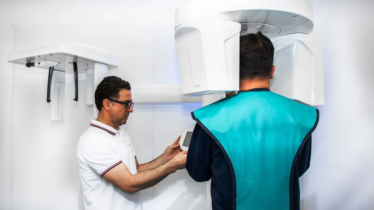
In four steps
- 01Clarifying the need
- 02Image in seconds
- 03Analysis on screen
- 04Discussion with you
FAQ
For new patients we take a panoramic and a bitewing X-ray, supplemented by a small single-tooth X-ray if needed. After that, a bitewing X-ray follows once a year to check for decay, and a panoramic X-ray every two years. Regular checks matter because some processes, including malignant ones, would otherwise go undetected.
It is small: according to model calculations, four small dental X-rays and one panoramic image carry the same risk as smoking one cigarette or a flight of over 9,600 km. Digital X-rays also reduce the radiation dose considerably, and the image appears on screen within seconds.
It gives an overview of all your teeth and the jawbones: the position of the teeth, the bone substance and any bone loss, and the quality of previous treatment. Remaining root fragments, changes in the maxillary sinus, decay and diseases of the bone and tooth roots also become visible.
A detailed image of individual teeth or areas of teeth. We take one when damage has been found or is suspected, for example a focus of pus (granuloma) or periodontitis, so that we can assess it precisely.
Yes, X-rays are particularly important for children. They reveal decay in baby teeth that can spread to the permanent teeth, decay in permanent teeth, misaligned teeth and missing or extra teeth.
Before implants, to measure the width and height of the available bone, and before surgical procedures such as wisdom tooth removal or apicoectomies. It also allows sources of inflammation and fractures of teeth and bone in the mouth, jaw and face to be diagnosed very precisely.
The image itself takes seconds; with preparation and discussion allow 5 to 15 minutes.
Usually not. Where necessary, we use local anaesthesia. For anxious patients we also offer treatment under general anaesthesia.
In Switzerland, dental treatment is usually not covered by basic health insurance. Many supplementary insurance plans contribute – we will gladly prepare a cost estimate for you to submit.
Usually within the same week – and on the same day if you are in pain. We offer emergency treatment Monday to Saturday until 21:00.